Mapping Aortic Valve Cells Using the FLUOVIEW FV3000 Microscope
The valves of a healthy adult heart allow blood to flow to the lungs and throughout the body. However, the structure and function of the aortic valve are vulnerable to environmental stress. Understanding the cellular composition of the aortic valve is critical for the treatment of aortic valvular diseases, such as bicuspid aortic valve calcification and leaflet degradation.
By using the FLUOVIEW FV3000 confocal microscope, we successfully confirmed the contribution of Tie2 positive cells to the aortic valve identified by lineage tracing technology.
Easily Perform Macro-to-Micro Imaging of Murine Aortic Valve Cells
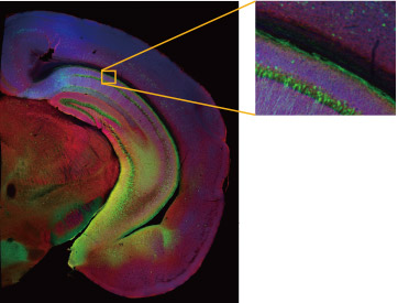
Thanks to the 1.25X macro imaging objective, we could easily and quickly acquire a large overview of an aortic valve slice with a single image. By simply dragging the mouse on the overview image, the FV3000 confocal microscope’s macro-to-micro function enabled us to quickly locate target cells and then observe the fine structures in a region of interest using a high-resolution objective lens.
Video: Macro-to-micro overview of the expression pattern of Tie2 in the aortic valve of a mouse. Tie2 positive cells are labeled with EGFP (green), and non-endothelial cells are labeled with td Tomato (red).
Imaging equipment
Microscope: FV3000 confocal microscope with a motorized stage
Objective: 1.25X, 10X, and 40X objective (PLAPON1.25X, UPLSAPO10X2, and UPLSAPO40X2)
How the FV3000 Confocal Microscope Facilitated Our Experiment
Macro-to-Micro ObservationThe FV3000 microscope’s macro-to-micro function provides a roadmap for data acquisition, enabling you to see data in context and easily locate regions of interest for higher resolution imaging. |
|
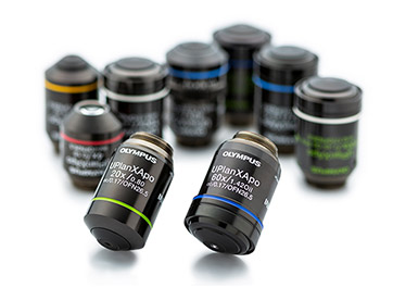
Extensive Lineup of Olympus ObjectivesOlympus offers a wide range of objectives. The FV3000 optical light path supports objectives from 1.25X to 150X and acquires images of samples from large tissues to small organisms. |
|
Acknowledgments
This application note was prepared with the help of Dr. Wenli Fan, State Key Laboratory of Pharmaceutical Biotechnology, Department of Cardiology, Nanjing Drum Tower Hospital, The Affiliated Hospital of Nanjing University.
References
- Feng, Q. et al. PDK1 regulates vascular remodeling and promotes epithelial-mesenchymal transition in cardiac development. Mol Cell Biol 30, 3711-3721, doi:10.1128/MCB.00420-10 (2010).
- Di, R., Yang, Z., Xu, P. & Xu, Y. Silencing PDK1 limits hypoxia-induced pulmonary arterial hypertension in mice via the Akt/p70S6K signaling pathway. Exp Ther Med 18, 699-704, doi:10.3892/etm.2019.7627 (2019).
Products Related to This Application
was successfully added to your bookmarks
Maximum Compare Limit of 5 Items
Please adjust your selection to be no more than 5 items to compare at once
Not Available in Your Country
Sorry, this page is not
available in your country.

