The new year is off to a running start, and while winter is in the air in North America, plants are still thriving on our Instagram feed. This month’s top images include some close-up views of cyanobacteria, plants, and flowers, making us anxious for spring to arrive.

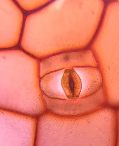
This image shows a stoma, a tiny pore on the surface of a leaf, stem, or other plant part that is used for gas exchange. Most leaves are covered with stomata, which allow the plants to take in carbon dioxide for use in photosynthesis. The resulting oxygen is released back through the stoma.
2019 Image of the Year submission. Captured on an Olympus CX23 microscope. Image courtesy of Eva Petrovova.

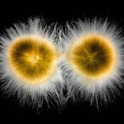
Double trouble! This photograph shows two colonies of cyanobacteria, Gloeotrichia echinulate. A type of cynobacterium that’s commonly found in freshwater ponds and lakes, it is infamous for forming dense blooms in nutrient-rich lakes. The cells can produce microcystins (liver toxins) and lipopolysaccharides (skin irritants) that can be harmful or even deadly. There are many records of livestock, dogs, and humans suffering liver damage through ingesting contaminated water.
This sample was collected in a local marina that suffered from an algae bloom. It was photographed using darkfield illumination with space between the slide and the cover glass to avoid it being squeezed too much to preserve its ball shape.
Description and image courtesy of Håkan Kvarnström.


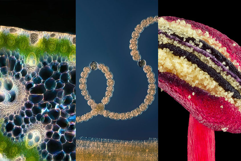
These images show a sample of bullrush, the common name for several grass-like wetland plants. This bullrush sample was collected from the banks of a pond in the National Botanic Gardens in Dublin, Ireland. Here, we can see a close-up look at a 20-micron section of the plant.
Captured using darkfield microscopy. Images courtesy of Karl Gaff.

This close-up of a flower stamen shows the anther of a Callistemon flower full of pollen. Callistemon is a genus of shrubs in the family Myrtaceae, endemic to Australia but widely cultivated in many other regions. They are often referred to as “bottlebrushes” due to the brush-like appearance of the flowers.
2019 Image of the Year submission. Image courtesy of Leonardo Capradossi.

There was a time when cyanobacteria dominated the Earth. Cyanobacteria have been present on Earth for as long as 4 billion years. At that time, there was almost no oxygen in the air. Over time, cyanobacteria developed the unique ability to take energy from the sunlight through photosynthesis and use it to make sugars out of water and carbon dioxide. Oxygen was the waste product that eventually led to the oxidization of the Earth's atmosphere, opening up conditions for new types of life, like animals. The other significant contribution of the cyanobacteria is the origin of plants. The chloroplast plants use to make food is actually a cyanobacterium living within the plant's cells. At some point in the past, cyanobacteria managed to enter other eukaryote cells, making food for the eukaryote host in return for a home. Still, cyanobacteria account for approximately 20 to 30 percent of the photosynthesis on the planet today and continue to play an important role in the composition of the atmosphere.
Specimen: A cyanobacterium called Anabaena. The bottom yellow part is another species of cyanobacteria, most likely Aphanizomenon. Captured in Lake Mälaren in Stockholm, Sweden.
2019 Image of the Year submission. Description and image courtesy of Håkan Kvarnström.
One of our favorite parts of the week is Waterbear Wednesday. In the most liked video this month, this cute tardigrade stops eating to say "hello" to the camera.
Approximately half a millimeter in length, the tardigrade’s complex mouth parts are visible here. This magnification also allows us to see its black eyes, which are a single cell. The circles within the body are also cells. This one has laid three eggs and is seen eating the microbial mat it was collected from (within a freshwater Florida spring at 4 m (13 ft) deep).
Captured at 400x magnification. Video courtesy of Hunter Hines.
To see more images like these, be sure to follow us on Instagram at @olympuslifescience!
Interested in sharing your own images? Visit our image submission site.
Related Content
Microscope Camera Best Practices for Fluorescence Imaging

